
The Mouth as a Diagnostic Mirror: Understanding Oral Mucosal Pathology
The oral cavity is a highly sensitive environment, often serving as a diagnostic mirror that reflects both localized dental health and systemic physical conditions. The delicate soft tissues lining the mouth—such as the tongue, palate, inner cheeks, and gums—are vulnerable to many different conditions. These mucosal changes, known clinically as oral lesions, can range from simple, self-healing benign mouth ulcers to persistent, pre-cancerous anomalies and aggressive, life-threatening malignancies.
Because early intervention is absolutely vital, identifying and diagnosing these tissue changes early is crucial. At V.I.P Dental Care, Varanasi, under the expert direction of Dr. V.R. Mishra, B.D.S., M.U.H.S. (Mumbai), we specialize in advanced oral mucosal screenings and early intervention. Because our Sigra branch is an Approved Tobacco Intervention Initiative (TII) Center, we place a high clinical focus on thorough soft tissue evaluations, low-pain biopsies, and patient-focused preventive care across our modern locations in Sigra, Kamachha, and Shivpur.
1. The Biology of the Oral Mucosa: How Tissue Changes Occur
The oral cavity is lined by a specialized, protective layer called stratified squamous epithelium, supported by a rich layer of connective tissue. This delicate tissue is constantly exposed to mechanical force from chewing, thermal changes from hot or cold foods, and chemical exposure from habits like smoking or using chewing tobacco, paan, and gutka.
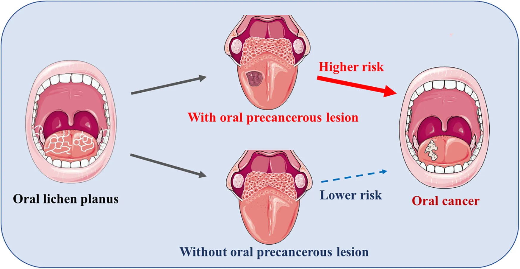
When subjected to chronic chemical or physical stress, the mucosal cells attempt to protect themselves by thickening their outer keratin layer, a process known as hyperkeratosis. If this irritation continues, the cells can gradually begin to develop abnormally, a condition called epithelial dysplasia. Left unmanaged, severe dysplasia can progress to carcinoma in situ and eventually develop into invasive oral cancer. Identifying these changes at the early, pre-cancerous stage is essential for successful, conservative management and recovery.
2. Identifying the Major Categories of Oral Lesions
Oral lesions are clinically classified based on their appearance, texture, and underlying causes. We routinely evaluate and manage several major categories of soft tissue conditions:
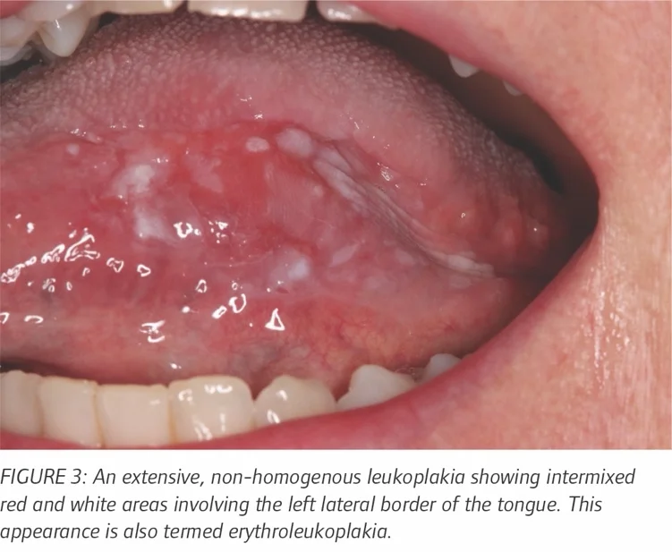
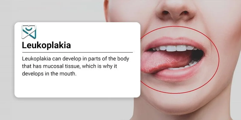
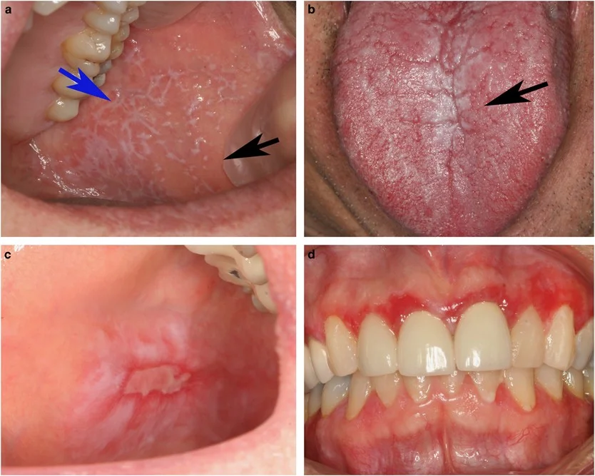
White Lesions (Leukoplakia & Lichen Planus): Leukoplakia presents as firm, raised white patches on the gums or inner cheeks that cannot be scraped away. Oral Lichen Planus, an autoimmune condition, often appears as a lacy network of fine white lines (Wickham's striae) or sensitive, red, and eroded areas on the delicate tissues of the mouth.
Red Lesions (Erythroplakia): Erythroplakias are velvety, red patches that develop on the soft tissues of the mouth. Because red lesions have an exceptionally high rate of pre-cancerous or malignant changes, they require immediate clinical evaluation, monitoring, and biopsy.
Ulcerative Lesions (Aphthous & Traumatic Ulcers): Canker sores (aphthous ulcers) are painful, self-healing lesions triggered by stress, dietary sensitivities, or minor tissue irritation. Traumatic ulcers are caused by physical irritation, such as a sharp tooth cusp, broken restoration, or ill-fitting partial dentures.
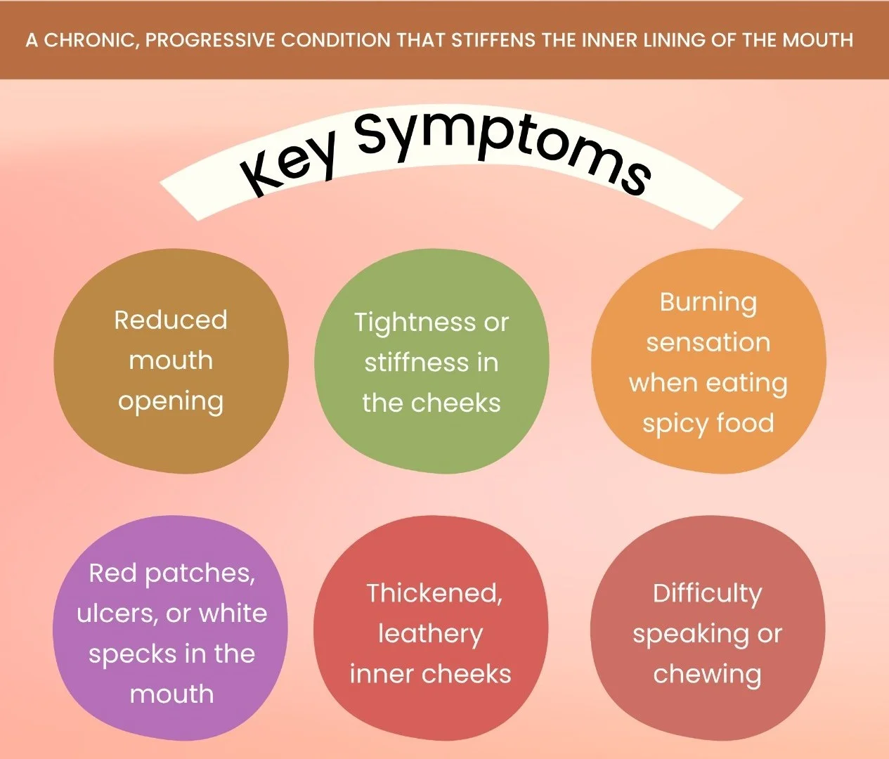
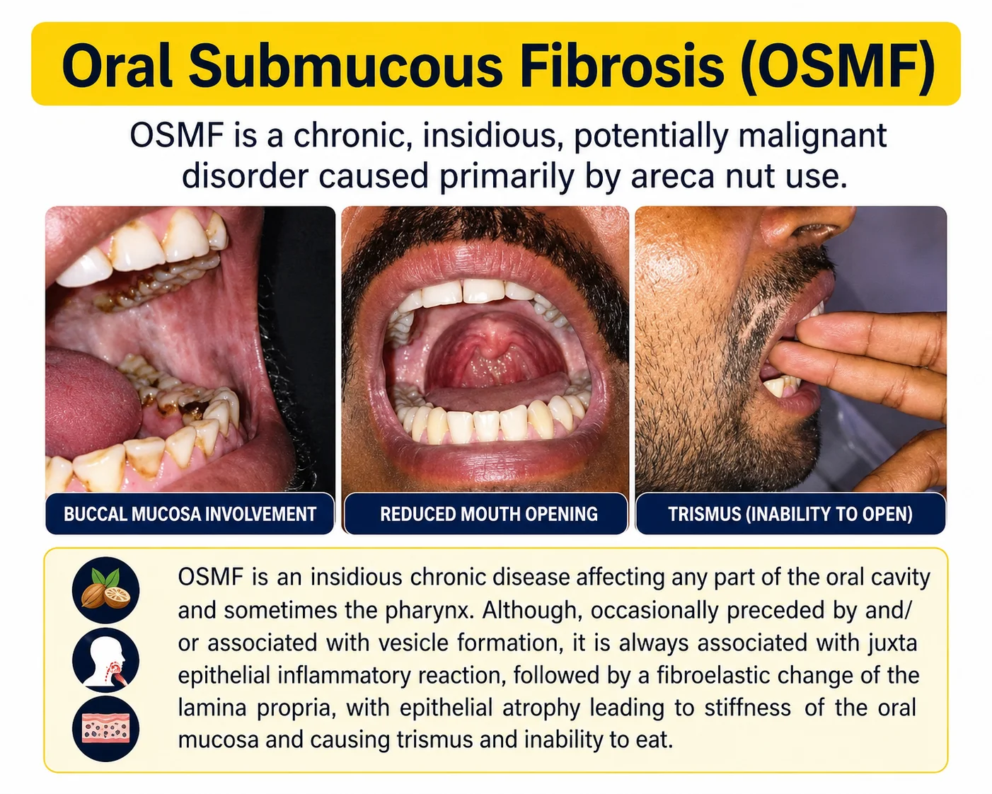
Oral Submucous Fibrosis (OSMF): This chronic, progressive scarring condition is primarily caused by chewing betel nut, gutka, and zarda. The toxic compounds cause dense, fibrous bands to develop in the cheeks, leading to progressive jaw stiffness (trismus) and difficulty opening the mouth, which requires immediate medical intervention.
3. Clinical Diagnostic Protocols and Biopsy Methods
When evaluating a persistent or unusual oral sore, we follow a precise diagnostic protocol to ensure an accurate, reliable diagnosis:
First, we perform a detailed visual and physical exam under high-powered surgical lights. If a lesion has been present for longer than two weeks without a clear cause of physical trauma, further investigation is necessary to ensure your safety.
We may recommend a low-pain diagnostic biopsy, which is the only definitive way to identify the cellular structure of the tissue. Depending on the size of the lesion, we perform either an incisional biopsy (taking a small sample of a larger area) or an excisional biopsy (removing a small, localized lesion completely). Both procedures are performed gently under local anesthesia, ensuring a comfortable, pain-free experience.
4. Tobacco, Gutka, and Varanasi's Oral Health Risk Profile
Varanasi and the broader Purvanchal region face unique oral health challenges. The widespread use of chewing tobacco, paan, gutka, and smoking bidis or cigarettes exposes oral tissues to a continuous stream of toxic, cancer-causing chemicals, significantly increasing the risk of oral cancer.
To help address this local health concern, our Sigra branch is an Approved Tobacco Intervention Initiative (TII) Center. This special accreditation allows our clinics to provide professional soft tissue screenings, detailed dependency evaluations, and customized cessation counseling. By combining meticulous oral screenings with supportive behavioral counseling, we help our patients protect their health and prevent the development of pre-cancerous lesions.
5. Medical Management and Rehabilitation of Oral Lesions
The treatment of an oral lesion depends entirely on its underlying cause and biopsy results. For autoimmune or inflammatory conditions like Lichen Planus, we provide customized, topical medical therapies to soothe discomfort. For fungal or viral infections, we prescribe targeted medications to clear the condition quickly.
For progressive pre-cancerous conditions like OSMF, we offer comprehensive treatment plans. This includes using rich antioxidant therapies (such as Lycopene), localized medication injections, and targeted physical exercises to improve jaw movement. Once the tissues are stable, we can also perform advanced dental cleanings (Teeth Cleaning & Scaling) to remove stubborn tobacco stains, helping you achieve a clean, healthy, and beautiful smile.
Prioritize Your Oral Mucosal Health Today
Widespread tobacco use, persistent mouth sores, or joint stiffness should never be ignored. With low-exposure digital diagnostics, meticulous soft tissue screenings, and experienced surgical methods, our clinics are equipped to provide the precise, high-quality care needed to support your health.
Take charge of your health. Complete our scheduling form below to request an oral lesion consultation with Dr. V.R. Mishra.