Varanasi's Premier Maxillofacial Trauma Centre: Restoring Form, Function, and Symmetry
Maxillofacial skeletal injuries—particularly mandibular (lower jaw) and maxillary (upper jaw) fractures—present complex anatomical challenges that extend far beyond general dentistry. These critical injuries often result from road traffic accidents, physical impact, sports collisions, or accidental falls. Because of the jaws' central role in facial structure, breathing, chewing, and speaking, a fracture demands immediate emergency evaluation, absolute diagnostic precision, and highly specialized stabilization.
At V.I.P Dental Care, Varanasi, we have established our reputation as a trusted Dental Maxillofacial Trauma Centre. Guided by the surgical expertise of Dr. V.R. Mishra, B.D.S., M.U.H.S. (Mumbai), we provide 24/7 emergency triage, advanced 3D diagnostics, and sophisticated surgical solutions to restore your facial symmetry and function. Our specialized trauma services are offered across our advanced clinics in Sigra, Kamachha, and Shivpur, ensuring rapid and comprehensive care when you need it most.
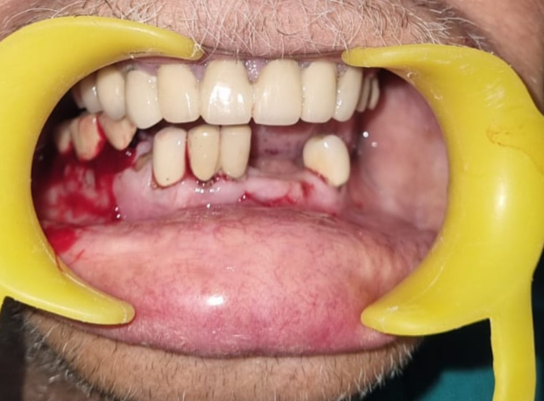

1. The Anatomy of Maxillofacial Trauma: Understanding Fracture Patterns
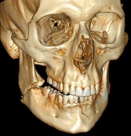
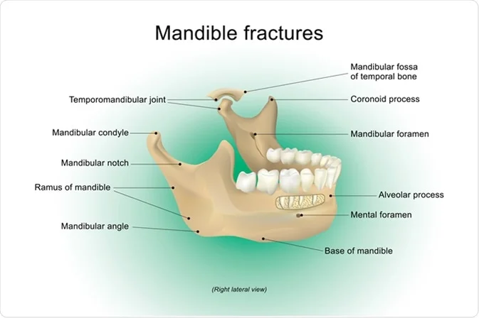
The human mandible is a complex, U-shaped bone that is the only mobile skeleton of the face. It is divided anatomically into several key segments: the symphysis, parasymphysis, body, angle, ramus, and the delicate condylar and coronoid processes. Understanding the unique structure of the jaw is essential, as the mandible’s dual-joint architecture (TMJs) means that a direct blow to one side of the jaw frequently causes an indirect fracture on the opposite side.
Maxillofacial fractures are clinically classified based on their location, complexity, and displacement:
- Simple (Closed) Fractures: The bone is fractured but remains contained within the overlying soft tissues, without tearing the gums or skin.
- Compound (Open) Fractures: The fracture line extends through the periodontal tissues or skin, creating a direct pathway for bacteria from saliva to enter the bone, significantly increasing the risk of infection.
- Comminuted Fractures: The force of the impact has shattered or crushed the bone into multiple small fragments, requiring detailed surgical stabilization.
- Condylar Fractures: Fractures occurring at the neck of the jaw joint. These require precise alignment to protect joint movement and prevent long-term complications like TMJ joint stiffness.
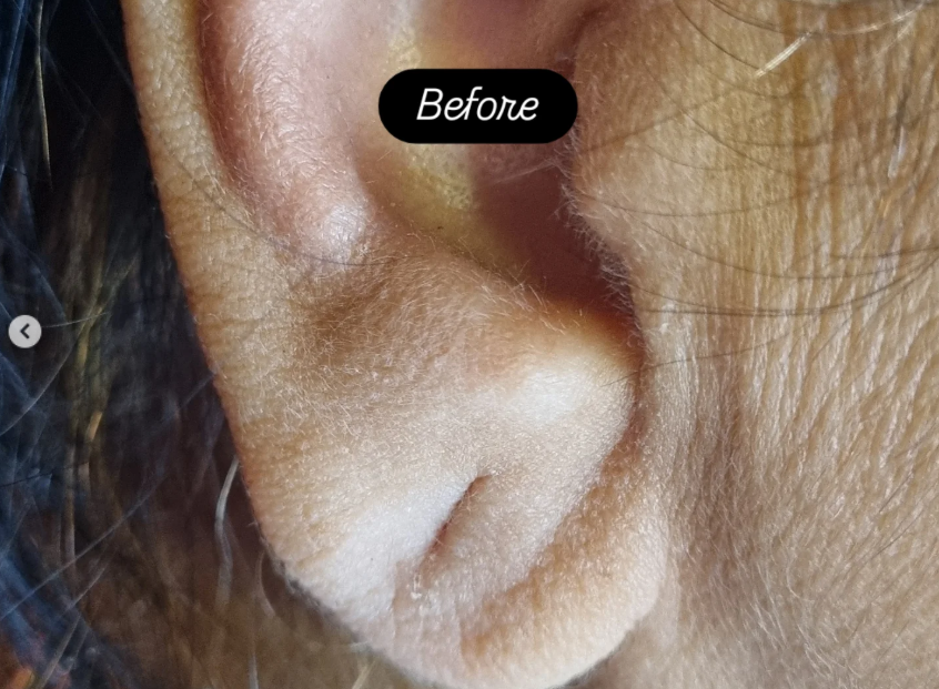
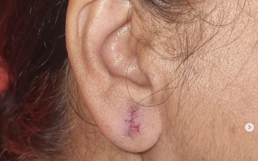
2. Emergency Diagnostics: Airway, Occlusion, and Advanced Imaging
In any maxillofacial trauma case, our immediate clinical priority is to secure the patient's airway and control bleeding. Once the patient is stable, we perform a thorough physical and radiographic evaluation. We check for key clinical signs of a jaw fracture, including an abnormal bite alignment (malocclusion), structural step deformities along the jawline, limited jaw opening, and potential numbness in the lower lip, which can indicate damage to the inferior alveolar nerve.
Advanced imaging is essential for planning treatment. We use low-radiation Orthopantomograms (OPGs) to provide an immediate panoramic baseline view of both jaws. In complex or multiple fracture cases, we utilize 3D Cone Beam Computed Tomography (CBCT) to map the injuries with millimeter precision, allowing us to plan the exact placement of surgical plates and screws with confidence.
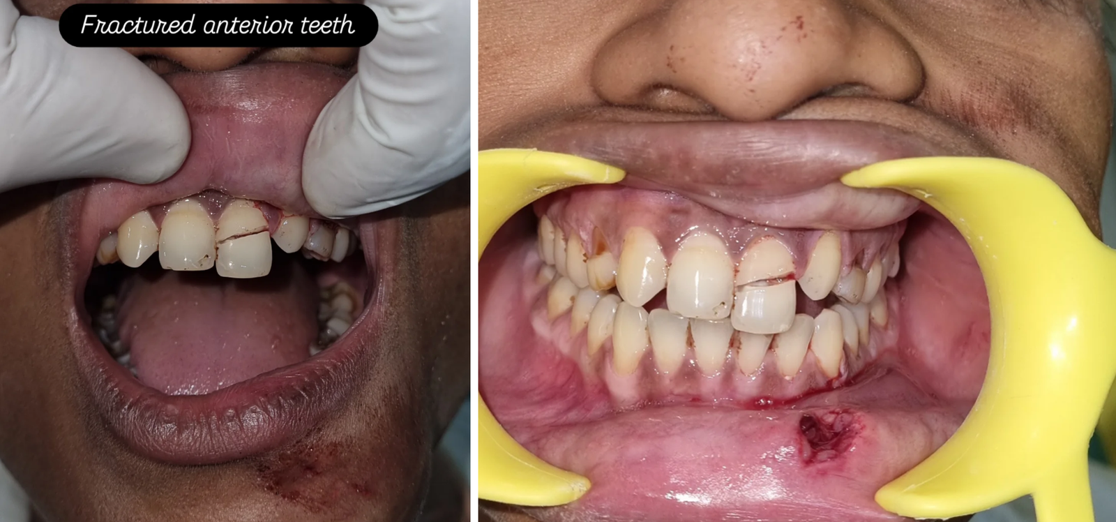
3. Non-Surgical Treatment: Intermaxillary Fixation (IMF)
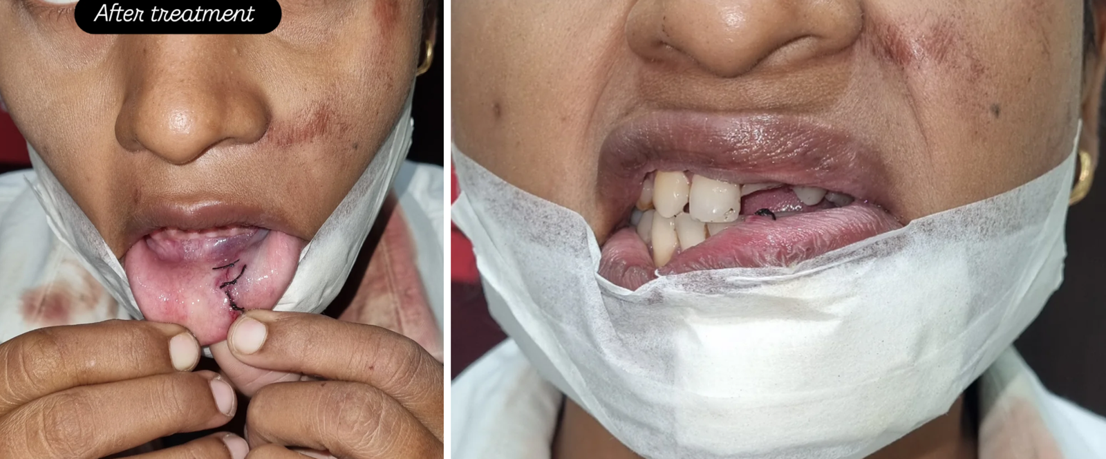
Not all jaw fractures require open surgery. If a fracture is stable, clean, and has not shifted out of position (undisplaced), we can manage it using conservative, non-surgical stabilization. This process, known as Intermaxillary Fixation (IMF) or closed reduction, focuses on alignment without incisions.
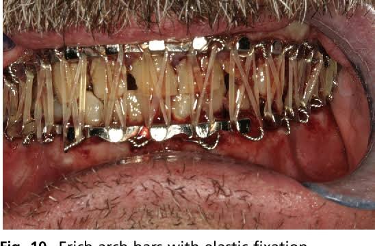
During closed reduction, we apply specialized metallic wires or Erich arch bars directly to the upper and lower dental arches. We then use fine wires or medical-grade elastics to align the teeth into their natural bite relationship and temporarily lock the jaws closed.
This stabilization is maintained for 4 to 6 weeks, holding the bone fragments perfectly still so they can heal. Because the jaws are locked closed, we provide patients with comprehensive nutritional guidelines and detailed home-care protocols to ensure they receive proper liquid nutrition and maintain excellent oral hygiene throughout their recovery.
4. Open Reduction & Internal Fixation (ORIF): Titanium Microplating
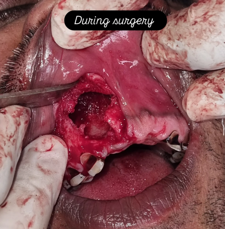
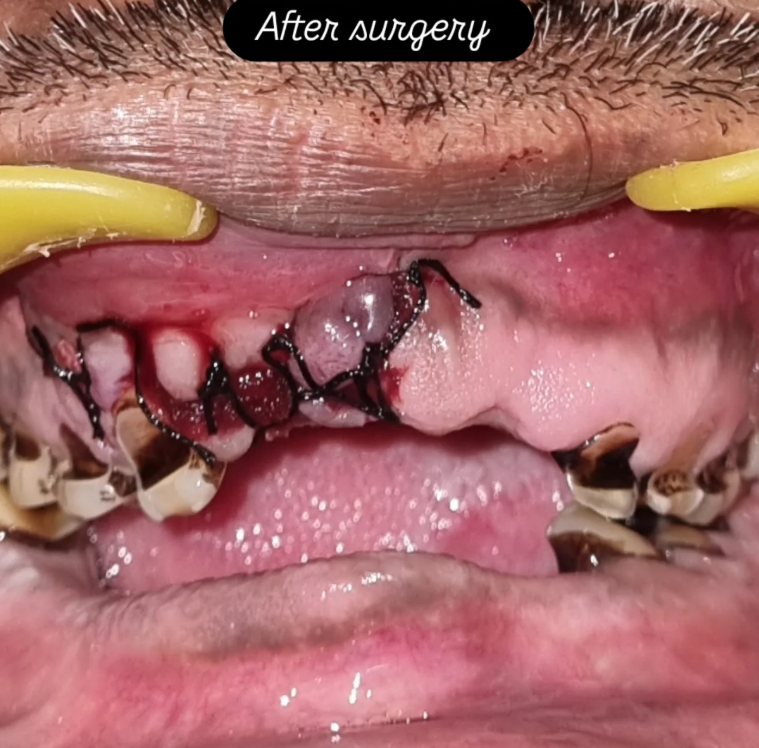
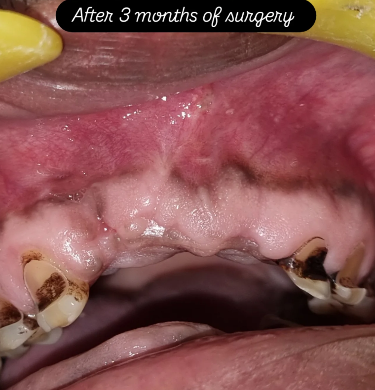
For displaced, unstable, or open compound fractures, surgery is necessary to ensure proper healing. Open Reduction and Internal Fixation (ORIF) is the gold standard for treating complex jaw fractures, allowing us to align and secure the bone fragments directly.
To avoid visible facial scars, we access the fractured bone using conservative, internal incisions made inside the mouth. Once the bone fragments are aligned, we use biocompatible titanium microplates and monocortical screws (typically 1.5mm to 2.0mm systems) to secure them across the fracture lines.
This rigid internal fixation provides immediate, stable support, allowing patients to gently move their jaws and return to a soft diet shortly after surgery. By reducing the need for long-term jaw wiring, ORIF helps prevent joint stiffness and muscle weakness, supporting a faster and more comfortable recovery.
5. Post-Trauma Rehabilitation and Complication Management
Our commitment to your recovery extends well beyond the operating room. Reconstructing a fractured jaw is a detailed process, and proper follow-up care is essential to ensure long-term success. We closely monitor your healing to prevent potential complications:
Bite Alignment Check (Occlusal Adjustments): We perform precise post-operative assessments to ensure your teeth meet together comfortably, preserving your natural bite and protecting your teeth from uneven wear.
Nerve Function Monitoring: Jaw trauma can sometimes stretch or bruise nearby nerves, causing temporary numbness. We closely track your sensory recovery and provide supportive therapies to help restore full sensation.
Temporomandibular Joint (TMJ) Therapy: Following periods of jaw stabilization, some joint stiffness is normal. We guide you through gentle, targeted exercises to restore a natural, comfortable range of movement.
Post-Surgical Dental Restoration: Once the jawbone has fully healed, our clinical team can address any damaged teeth with advanced treatments like zirconia Dental Crowns or dental implants, fully restoring both your smile's function and appearance.
6. Advanced Craniofacial Rehabilitation: Custom Auricular Prosthesis
Severe maxillofacial trauma often extends beyond skeletal fractures to involve significant soft tissue loss, including damage to or avulsion of the outer ear (auricle). Whether caused by road traffic accidents, burns, or congenital anomalies like microtia, the loss of an ear has profound psychological and functional impacts. At V.I.P Dental Care, our maxillofacial reconstructive services include the design and fabrication of custom auricular prostheses.
An auricular prosthesis is a customized, medical-grade silicone ear designed to match the unique anatomy, contours, and skin tone of the patient’s opposite, healthy ear. Dr. V.R. Mishra works closely with specialized laboratory partners to capture fine details, including natural skin variations and surface blood vessel patterns, ensuring a lifelike appearance. Depending on the patient's remaining bone and tissue structures, the prosthesis can be secured using medical-grade adhesives or anchored permanently using advanced osseointegrated craniofacial implants. Implant-retained ears use microscopic titanium clips or magnetic connectors to provide exceptional stability, letting patients return to their daily routines with confidence.
Trust Varanasi's Specialized Trauma Center
Maxillofacial skeletal injuries and soft tissue defects require immediate, specialized care to ensure proper healing. With advanced digital 3D scans, low-exposure OPG technology, and experienced surgical and prosthetic methods, our clinics are equipped to provide the precise, high-quality care needed to support your recovery.
Protect your long-term health. Complete our clinical intake form below to request a prioritized trauma or reconstructive consultation with Dr. V.R. Mishra.